Excitation sources
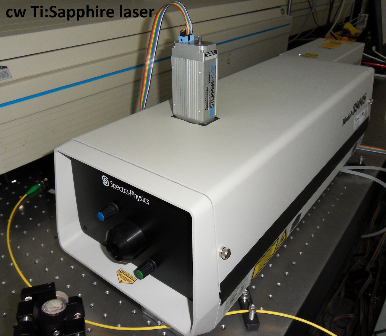
Optical Parametric Oscillator (OPO) pumped by a frequency tripled Nd:YAG laser. This OPO provides 10-ns pulses and it is tunable from 450 up to 1800 nm. Single-mode fibre-coupled lasers diodes at 1450, 1283, 1020, 980, 975, 808, 800, 794, and 690 nm for fluorescence microscopy. 532 nm solid state laser for confocal fluorescence microscopy. 50 mW 488 nm solid state laser for confocal fluorescence microscopy. 20 mW 405 nm solid state laser for confocal fluorescence microscopy. 10 W Fibre coupled diode laser at 808 nm. 25 W Fibre coupled diode laser at 980 nm. Ti:Sapphire laser providing 100-fs laser pulses at a repetition rate of 80 MHz. This laser is tunable from 750 up to 1000 nm. Continuous wave Ti:Sapphire Laser pumped by a 15 W 532 nm solid state laser. Tunable from 700 up to 1100 nm. Air-cooled Argon laser (300 mW) for micro-Raman measurements. 10 W Yb fibre laser operating at 1090 nm (continuous wave).

Microscopes
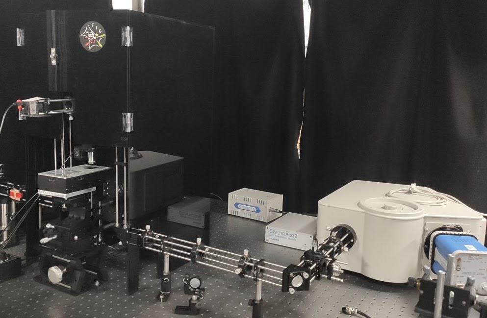
Home-made two-beam confocal fluorescence microscope for fluorescence imaging of living cells during laser treatment and fluorescence mapping. Home-made optical trapping motorized set-up coupled to the confocal fluorescence microscope for fluorescence imaging of optical traps. Home-made infrared and visible confocal microscope for hyperspectral imaging in the 400-1700 nm spectral range. Commercial dark field microscope (Nikon) with the possibility of spectral analysis of dark field image. Epi-fluorescence microscope excited by a UV lamp for fluorescence imaging of micro-fluidic devices and living cells. Commercial (Horiba) modular micro-Raman microscope. Optical coherence tomography setup for intravascular imaging (St. Jude Medical). Optical coherence tomography setup (Thorlabs). Optical system for dark field microscopy. Includes optical systems for the measurement of scattering spectra in the visible and infrared ranges (400-1700 nm).


Detection
Andor micro-monochromator coupled to an InGaAs CCD array (for spectral detection in the 700-1700 nm spectral range) and to an electron-multiplying CCD array (for imaging and spectral detection in the 400-1100 nm range). Photomultipliers for lifetime measurements covering the 400-1100 nm detection range. Andor micro-monochromator coupled to an infrared Hamamtsu photomultiplier for lifetime measurements in the 800-1700 nm spectral range. Thermal imaging camera (Flir). 0.5 M Horiba i320 monochromator. Working range 250-1800 nm. 0.25 M Horiba i320 monochromator. Working range 250-1800 nm. 2 silicon detection arrays for luminescence detection in the confocal fluorescence microscopes. Detection range from 400 up to 1100 nm. (Horiba) 1 InGaAs detection array for luminescence measurements in the confocal fluorescence microscopes. Detection range from 850 up to 1600 nm. Germanium detector for detection in the 800-1500 nm spectral range. 500 MHz digital oscilloscope. Fibre-coupled avalanche photo-detectors for short pulse measurements in the 800-1400 nm spectral range and in the 100 ps time scale. High-gain CCD camera for fluorescence imaging and laser beam analysis. InGaAs CCD camera (XEVA 17 from Xenics) for infrared fluorescence imaging in the 800-1700 nm spectral range. Auto-correlator for laser pulse analysis. Lambda 1050 UV/Vis/NIR spectrophotometer (PerkinElmer) working from 175 to 3300 nm.Synthesis and functionalization of nanoparticles
Our wet-chemistry laboratory is equipped with two fume hoods and all the necessary infrastructure/instrumentation for the synthesis and complete characterization of nanoparticles. The lab includes:
Schlenk lines Heating mantles with temperature controllers Heating plates with magnetic stirring Vortex Sonicators (bath and tip) Laboratory oven Malvern Zetasizer Ultra Heidolph G3 rotary evaporator Ortoalresa Biocen 22R centrifuge with controlled temperature HERMLE Z206S centrifuge Sonics Vibracell Ultrasonic Homogeneizer CEM Discover 2.0 Microwave ReactorAnimal imaging
Home-made chamber for small animal infrared imaging. Excitation at 808 nm and detection range spanning from 800 up to 1700 nm. Home-made chamber for small animal photothermal therapies with capability of thermal imaging with a 1-°C resolution. Laser speckle camera for subcutaneous flow measurements. Home-made chamber for two-dimensional fluorescence imaging of infarcted heart.We also work in close collaboration with the School of Medicine and the Department of Biology of UAM, through which we can access cell cultures and small animal facilities.
